knee and leg
About
knee bones
bones
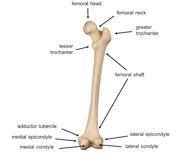
femur
The femur is the only bone in the thigh and the longest bone in the body. It acts as the site of origin and attachment of many muscles and ligaments, and can be divided into three areas; proximal, shaft and distal. In this article, we shall look at the anatomy of the femur – its attachments, bony landmarks and clinical correlations.
Learn More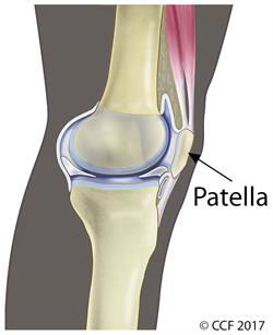
patella
The kneecap by another name, the patella is the small bone that is in the front of the knee The patella is a small bone located in front of the knee joint — where the thighbone (femur) and shinbone (tibia) meet. It protects the knee and connects the muscles in the front of the thigh to the tibia. The ends of the femur and the undersides of the patella are covered with a smooth substance called articular cartilage. This cartilage helps the bones glide easily along each other as you move your knee.
Learn More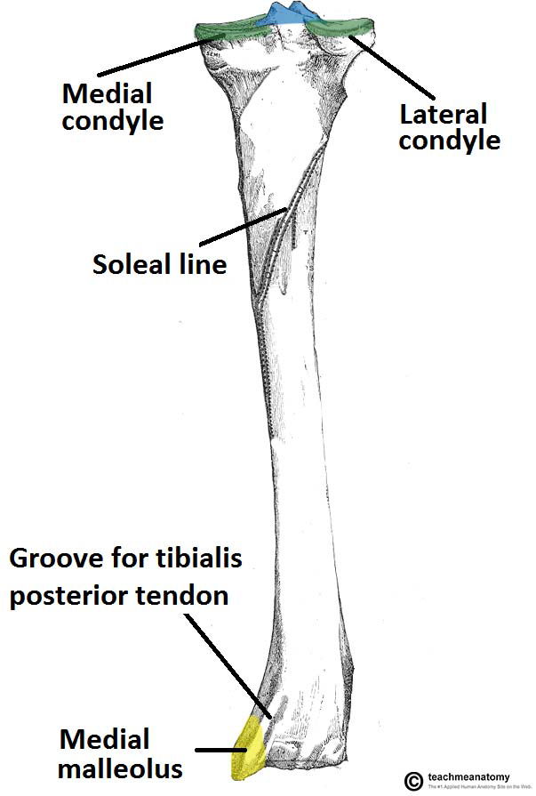
tibia
The tibia, sometimes known as the shin bone, is the larger and stronger of the two lower leg bones. It forms the knee joint with the femur and the ankle joint with the fibula and tarsus. Many powerful muscles that move the foot and lower leg are anchored to the tibia. The support and movement of the tibia is essential to many activities performed by the legs, including standing, walking, running, jumping and supporting the body’s weight.The tibia is located in the lower leg medial to the fibula, distal to the femur and proximal to the talus of the foot.
Learn More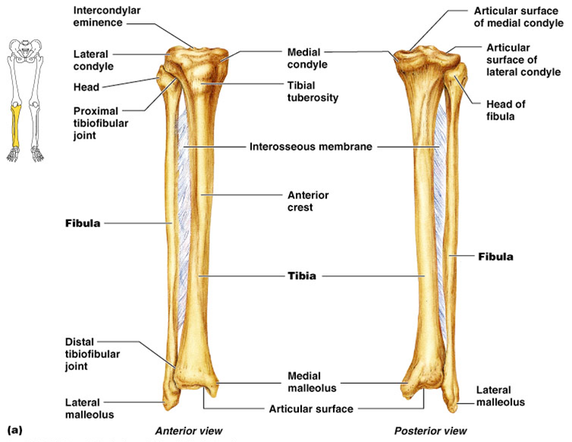
fibula
The fibula is the long, thin and lateral bone of the lower leg. It runs parallel to the tibia, or shin bone, and plays a significant role in stabilizing the ankle and supporting the muscles of the lower leg. It runs parallel to the tibia, or shin bone, and plays a significant role in stabilizing the ankle and supporting the muscles of the lower leg. Compared to the tibia, the fibula is about the same length, but is considerably thinner.
Learn Morebone marking
bone markings
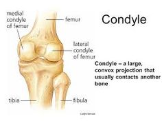
condyles
there are lateral and medial condyles on both the femur and the tibia. these serves as attachment points for muscles as well as the articulation point between the femur and the tibia
Learn More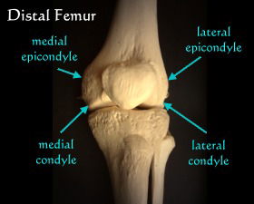
epicondyles
it is a bony protrusion that serves as attachment points. there are medical and lateral epicondyles on the femur
Learn More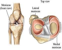
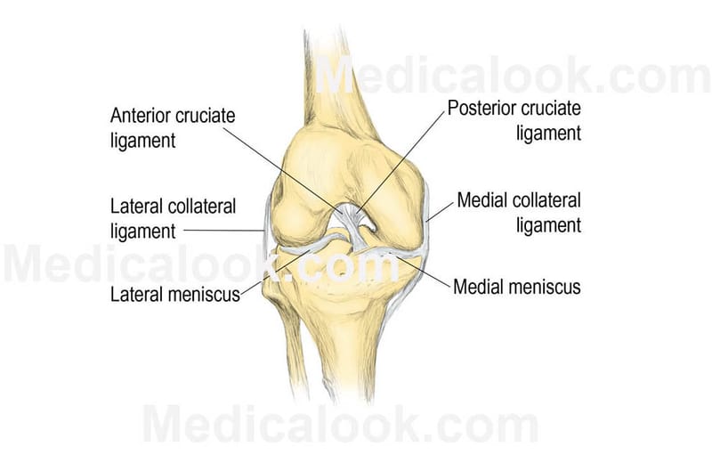
menisci
At knee joint the menisci plays a major role in congurency of the joint. Menisci forms the concavity in which the femoral condyles sits. Menisci rests between the thigh bone femur and the tibia and there are two knee joint ligaments. They are a type of cartilage in the joint. The rubbery texture of the menisci is due to their fibrocartilagenous structure. Their shape is maintained by the collagen within them. One meniscus is on the inner side of your knee--the medial meniscus. The other meniscus is on the outer side of your knee - the lateral meniscus.
Learn More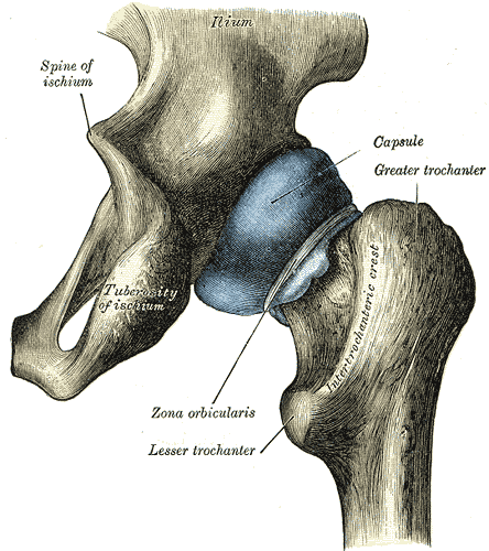
greater trochanter
The greater trochanter (great trochanter) of the femur is a large, irregular, quadrilateral eminence and a part of the skeletal system. It is directed lateral and medially and slightly posterior.
knee ligaments
ligaments
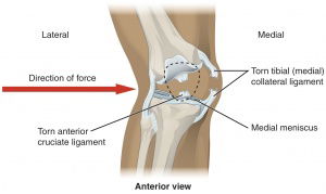
medial collateral ligament
The MCL is one of four major ligaments that supports the knee. This ligament is a strong broad band[1] found on the inner aspect of the knee joint and is the largest structure situated on the medial side.
Learn More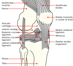
lateral collateral ligaments
The lateral collateral ligament (LCL) is one of four critical ligaments involved in stabilizing the knee joint. The medial collateral ligament, the anterior cruciate ligament and the posterior cruciate ligament are the other stabilizers of the knee.The lateral collateral ligament or fibular collateral ligament has its origin on the lateral epicondyle of the femur and runs to the fibular head.
Learn More
anterior cruciate ligament
The anterior cruciate ligament (ACL) is a band of dense connective tissue which courses from the femur to the tibia. The ACL is a key structure in the knee joint, as it resists anterior tibial translation and rotational loads.Arises from the posteromedial corner of medial aspect of lateral femoral condyle in the intercondylar notch[1]. This femoral attachment of ACL is on posterior part of medial surface of lateral condyle well posterior to longitudinal axis of the femoral shaft. The attachment is actually an interdigitation of collagen fibers & rigid bone thru transitional zone of fibrocartilage and mineralized fibrocartilage
Learn More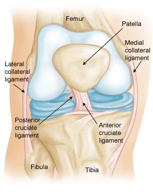
posterior cruciate ligament
The posterior cruciate ligament is located in the back of the knee. It is one of several ligaments that connect the femur (thighbone) to the tibia (shinbone). The posterior cruciate ligament keeps the tibia from moving backwards too far.
Learn Morejoints
tibiofemoral joint
The knee joint is one of the strongest and most important joints in the human body. It allows the lower leg to move relative to the thigh while supporting the body’s weight. Movements at the knee joint are essential to many everyday activities, including walking, running, sitting and standing.
Learn More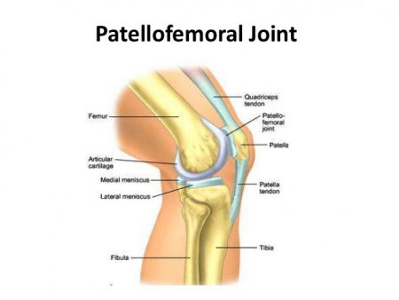
patellofemoral joint
Joint, patellofemoral: One of the knee joints. The knee has three parts. The thigh bone (femur) meets the large shin bone (tibia) forming the main knee joint. This joint has an inner (medial) and an outer (lateral) compartment. The kneecap (patella) and the femur form a third joint, called the patellofemoral joint.
Learn More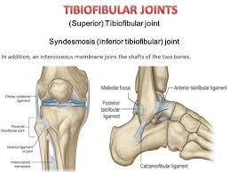
tibiobfbular (superior) joint
superior tibiofibular joint (also known as the proximal tibiofibular joint) is the joint between your tibia and fibula (lower leg bones) immediately below the outside of your knee. This joint is important in allowing twisting movements of the leg, as well as load transfer between your feet and the rest of your body.
Learn Moreanterior leg muscles
muscles and tendons
anterior muscles
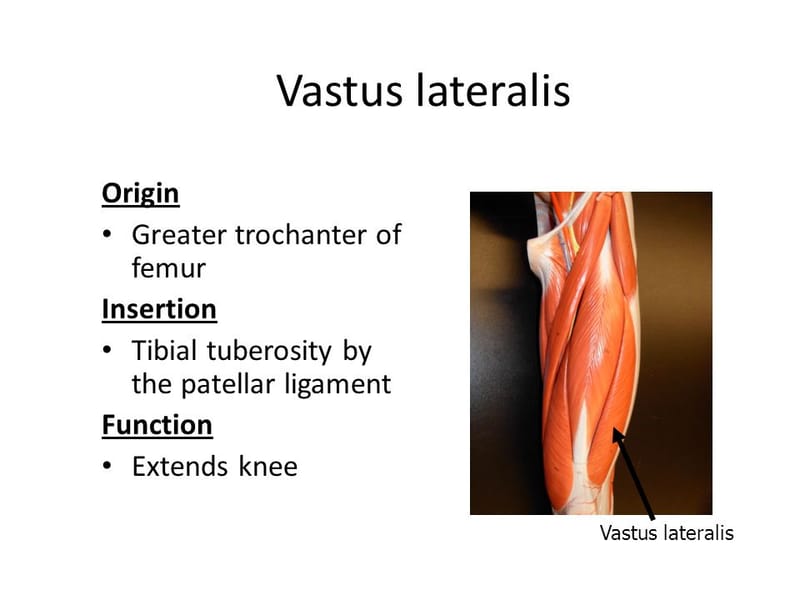
vastus lateralis
it covers the outer anterior aspect of the femur, arises chiefly from the femur, and inserts into the outer border of the patella by a flat tendon which blends with that of the other divisions of the muscle and sends an expansion to the capsule of the knee. The vastus lateralis muscle is located on the side of the thigh. Collectively, the quadriceps muscle is the largest in the human body and its purpose is to extend the knee. The specific task of the vastus lateralis muscle is to extend the lower leg and allow the body to rise up from a squatting position.
Learn More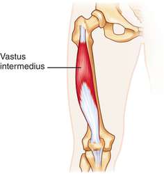
vastus intermedius
The vastus intermedius muscle is located along the upper portion of the femur, which is also known as the thighbone. This muscle covers the front part of the femur and the side of the femur. It is directly underneath and ends at the rectus femoris muscle, which is part of the quadriceps cluster of muscles.
Learn More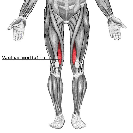
vastus medialis
vastus medialis muscle is a part of the quadriceps muscle group, located on the front of the thigh. It is the most medial, or inner, of the quadriceps muscles. It extends the entire length of the thigh. The portion of the muscle that is just above the knee is sometimes referred to as the vastus medialis obliquus, or VMO. This muscle is used to extend the leg at the knee and to stabilize the patella, which is also known as the kneecap.
Learn More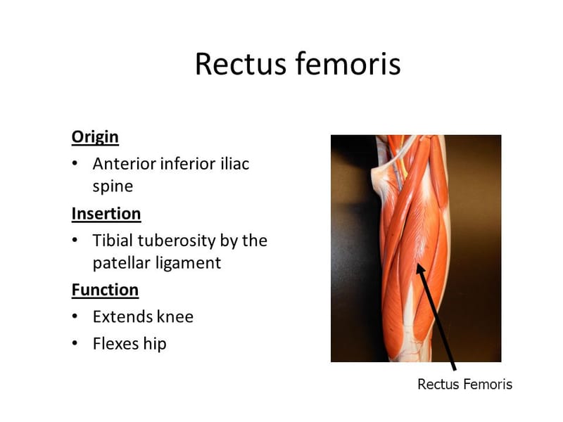
vastus femoris
it is the last of quadriceps muscles. origin- anterior inferior iliac spine insertion- tibial tuberosity by the patellar ligament function- extends knee flexes hip
Learn More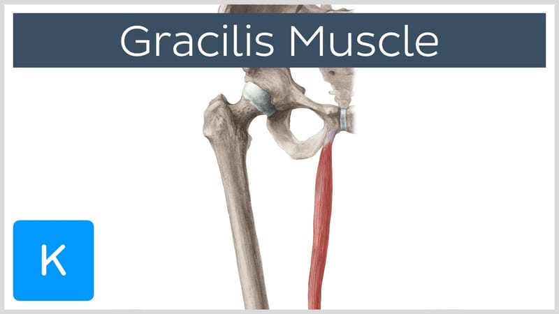
gracilis
The gracilis muscle is one of the muscles found in the groin.It starts at the external point of the ischiopubic ramus (on the pubic bone) and extends down to the upper medial (middle) shaft of the tibia, or shinbone. The gracilis is responsible for hip adduction and assists knee flexion
Learn More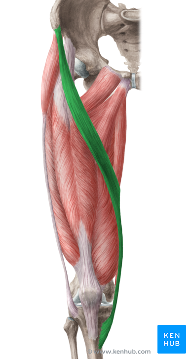
sartorius
Long and thin, the sartorius muscle spans the distance of the thigh. It originates at anterior superior iliac spine (a bony projection on the uppermost part of the pelvis) and travels to the upper shaft of the tibia, or shinbone. As such, the sartorius is the longest muscle in the human body.The muscle helps flex, adduct, and rotate the hip. In addition, it helps with the knee's flexion
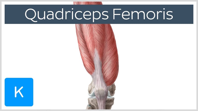
quadricep muscle
The Rectus femoris muscle has two origins at the anterior inferior iliac spine of the pelvis and the upper margin of the acetabulum. Distally its fibers end in the common insertion tendon (quadriceps tendon) Function: Extends the knee joint and stabilizes the patella.
adductors
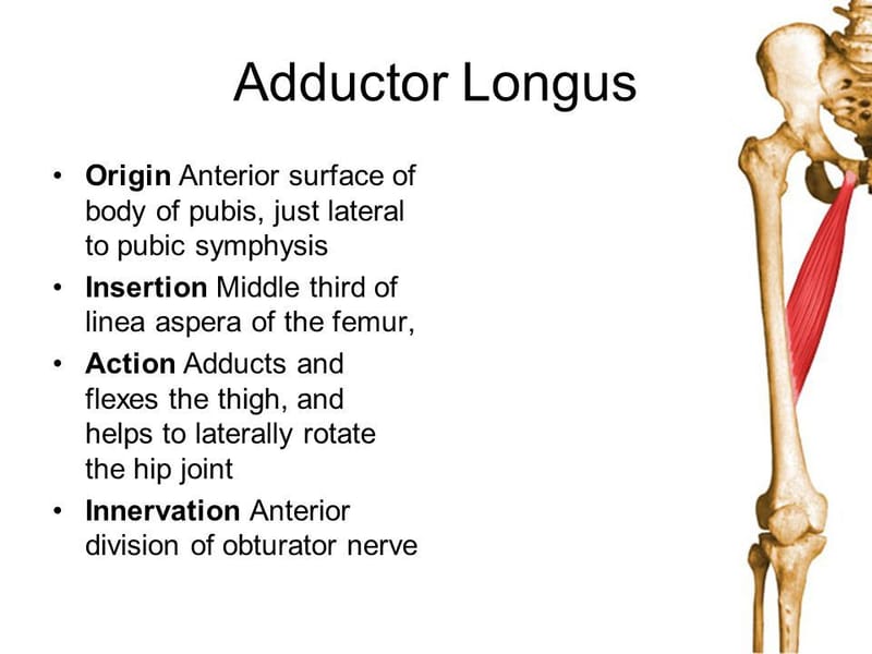
adductor longus
The adductor longus muscle is a hip abductor muscle located in the inner thigh. This muscle controls the thigh bone's ability to move inward and from side to side.The muscle originates in the superior aspect of the pubis, below the pubic tubercle. It inserts at the middle third of the linea aspera of the femur along the medial lip. It adducts and flexes the thigh at the hip. It also contributes to lateral and medial rotation of the thigh. All adductor muscles in the thighs pull the legs toward the middle when walking,
Learn More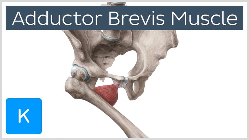
adductor brevis
The adductor brevis muscle helps to adduct the thigh at the hip joint. It can also flex and medially rotate the thigh. The adductor brevis muscle emerges from the body at the inferior ramus of the pubis. It inserts into the pectineal line and the middle of the linea aspera of the femur. The blood supply for this muscle originates from branches of the femoral and obturator arteries.
Learn More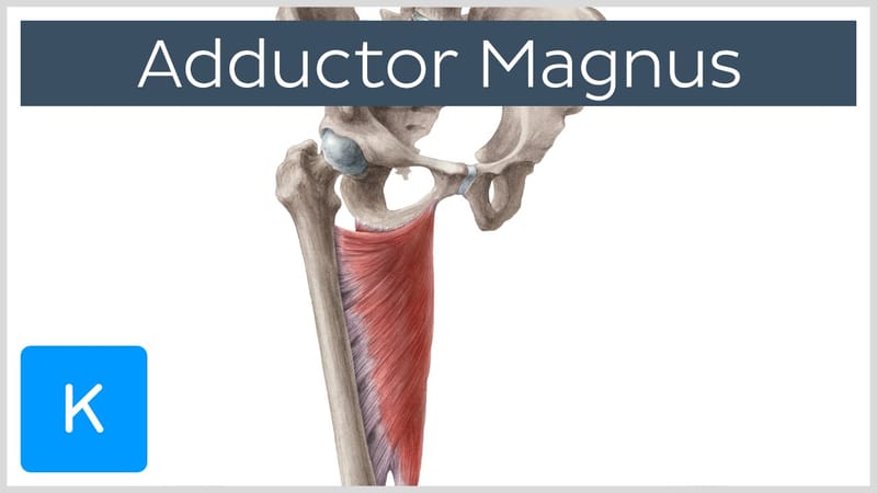
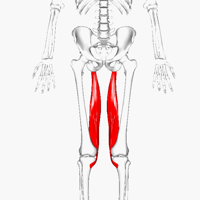
adductor magnus
On the medial side (closest to the middle) of the thigh, the adductor magnus muscle creates the shape of a large triangle. As an adductor, it contracts and pulls the hip towards the body's midline. This action is a fundamental part of walking, sprinting, and a variety of other bipedal motions. The muscle also extends the hip
Learn Moreposterior leg
semimembranosus
The semimembranosus muscle is one of the three muscles that make up the hamstring. It is found on the back of the thigh and runs from the base of the pelvis (specifically the tuberosity of the ischium) to the back of the tibia, one of the bones that make up the lower leg. The semimembranosus muscle is attached to the pelvis and tibia.The muscle has several functions, including enabling the leg to flex and rotate, and serving as a thigh extensor. The other two muscles that make up the hamstring are the semitendinosus and biceps femoris muscles.
Learn More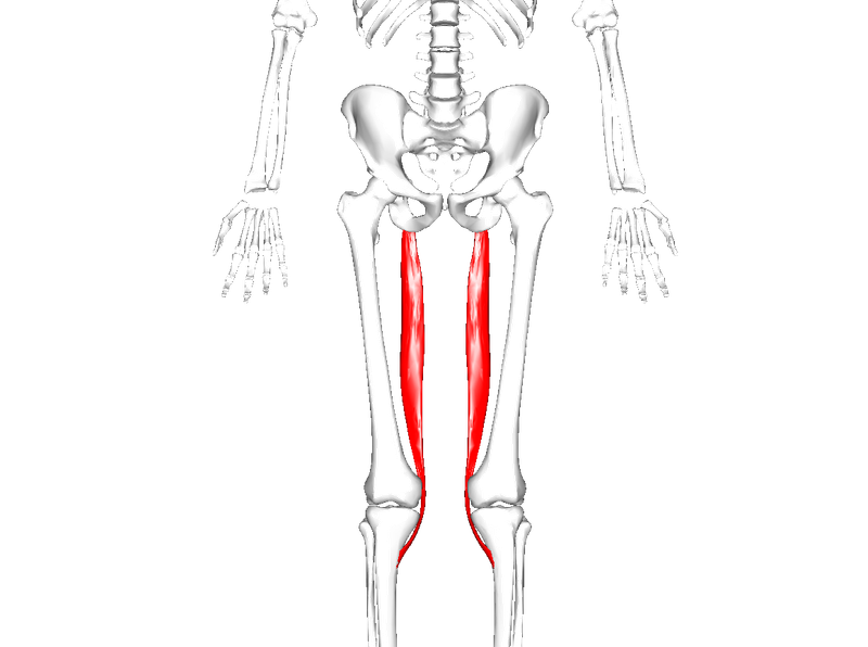
semitendinosus
The semitendinosus muscle is one of three hamstring muscles that are located at the back of the thigh. The other two are the semimembranosus muscle and the biceps femoris. The semitendinosus muscle lies between the other two. These three muscles work collectively to flex the knee and extend the hip.The semitendinosus muscle begins at the inner surface of the base of the pelvis (known as the tuberosity of the ischium) and the sacrotuberous ligament. It inserts at the medial tibial condyle.
Learn More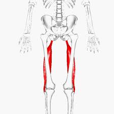
biceps femoris
The biceps femoris is a double-headed muscle located on the back of thigh. It consists of two parts: the long head, attached to the ischium (the lower and back part of the hip bone), and the short head, which is attached to the femur bone.The long head is a part of the hamstring muscle group that occupies the posterior section of the thigh. The hamstring muscles may be considered extensors of the thigh. The biceps femoris muscle is important for knee flexion, internal and external rotation, and hip extension.
Learn More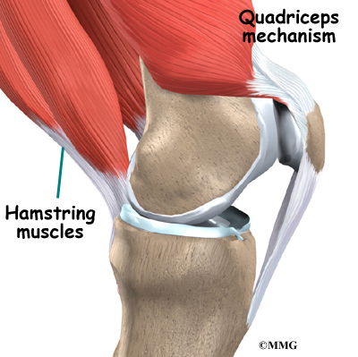
patella quadricep tendon
Quadriceps tendinitis (also spelled tendonitis) is a painful condition affecting the quadriceps tendon above the patella (kneecap). The quadriceps group of muscle (individually called the vastus lateralis, vastus intermedius, vastus medialis and the overlying rectus femoris) is the largest in the front of the thigh and is very important for stabilizing the leg. The quadriceps tendon connects the quadriceps to the tibia (shins) via the patellar tendon, which surrounds the knee cap itself. The movement of the knee known as the quadriceps mechanism involves the patella (knee cap), the patella tendon, the quadriceps and its tendon. They all work together to extend the leg to straight.
Learn More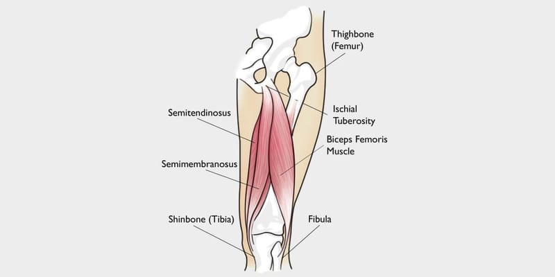
hamstring tendon
Hamstring tendonitis occurs when the soft tissues that connect the muscles of the back thigh to the pelvis, knee, and lower legs become inflamed. Tendonitis is often brought on by overuse and causes acute, or immediate, pain that decreases with rest and minor first aid. Most people can return to regular activity after a week or so. Full recovery typically involves rehabilitative exercises and takes several weeks.
Learn More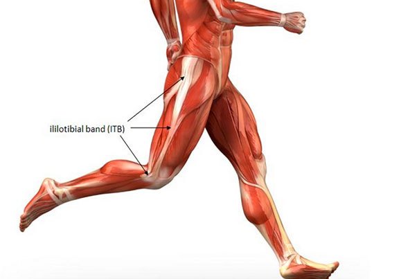
iliotibial band
The iliotibial tract or iliotibial band (IT band) is a longitudinal fibrous reinforcement of the fascia lata. The action of the ITB and its associated muscles is to extend, abduct, and laterally rotate the hip. In addition, the ITB contributes to lateral knee stabilization.
origin and insertion
anterior origin and insertion
posterior origin and insertion
injuries
injuries
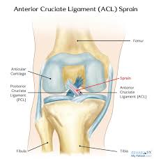
ACL SPRAIN
An ACL sprain is a tear to the anterior cruciate ligament in the knee which runs diagonally from the back of the femur (thigh bone) upwards and forwards to the front of the tibia (shin bone) and prevents the shin bone from moving excessively forward. It usually occurs as a result of either a twisting force being applied to the knee whilst the foot is firmly planted on the ground or twisting of the knee when landing from a jump. treatment- depends on severity
Learn More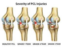
PCL sprain
The posterior cruciate ligament (PCL) is a ligament within the knee. Ligaments are tough bands of tissue that connect bones. The PCL -- similar to the anterior cruciate ligament (ACL) -- connects the thigh bone (femur) to your shin bone (tibia). Although it is larger and stronger than the ACL, the PCL can be torn. PCL injuries are often due to a blow to the knee while it's bent. Common causes include: Striking the knee against the dashboard during an auto accident Falling on the knee while it's bent Most people don't feel or hear a "popping" sensation in the knee after a PCL injury. This is more common with an injury to the ACL. treatment depends on the severity
Learn More
LCL sprain
The main cause of LCL injuries is direct-force trauma to the inside of the knee. This puts pressure on the outside of the knee and causes the LCL to stretch or tear. Symptoms of an LCL injury can be mild or severe, depending on the severity of the sprain or if it’s torn. If the ligament is mildly sprained, you may not have any symptoms at all. For a partial tear or complete tear of the ligament The treatment options for LCL injuries will depend on the severity of the injury
Learn More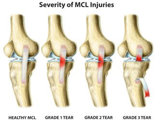
MCL sprain
it occus when knee is forced laterally swelling.bruising and discomfort are the symptoms treatment- rice to surgery depends.
Learn More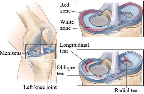
meniscus injuries
a meniscus tear can be painful and debilitating. Unfortunately, it's quite common. In fact, a meniscal tear is one of the most frequently occurring cartilage injuries of the knee.Meniscus tears are common in contact sports like football as well as noncontact sports requiring jumping and cutting such as volleyball and soccer. They can happen when a person changes direction suddenly while running, and often occur at the same time as other knee injuries, like an anterior cruciate ligament (ACL) injury. Meniscus tears are a special risk for older athletes since the meniscus weakens with age. More than 40% of people 65 or older have them. Treatment of a torn meniscus may include observation and physical therapy with muscle strengthening to stabilize the knee joint. When conservative measures are ineffective treatment may include surgery to repair or remove the damaged cartilage.
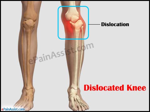
dislocation
A knee dislocation occurs when the bones that form the knee are out of place. A knee dislocation, more specifically, is when the bones of the leg (the tibia and fibula) are moved in relation to the bone in the thigh (femur).A knee dislocation occurs when the bones that form the knee are out of place. A knee dislocation, more specifically, is when the bones of the leg (the tibia and fibula) are moved in relation to the bone in the thigh (femur). Inappropriate or delayed treatment of a knee dislocation may result in loss of the leg.
fractures
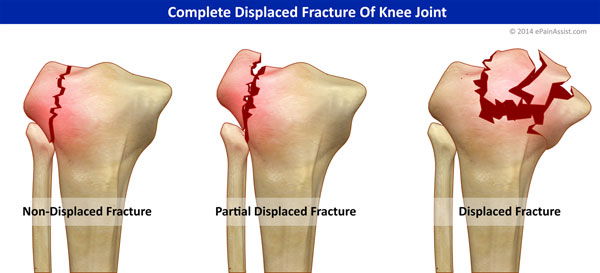
A kneecap fracture is a break or crack in the kneecap (patella). It may be just a small crack in the bone, or the bone may break into pieces or shatter. Some fractures may stick out through the skin. broken kneecap usually results from a fall onto your knee or a direct hit to the knee. Some kneecap fractures can happen when you are jumping or running. treatment- surgery
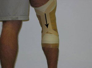
tape job
tape jobs
knee hyperextension
A hyperextended knee occurs when your knee is pushed past its normal range of motion from a straightened position
Learn More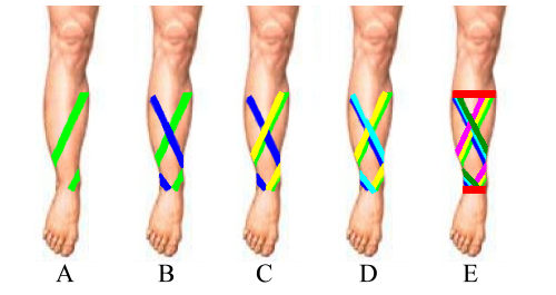
shin splint
acute pain in the shin and lower leg caused by prolonged running, typically on hard surfaces.
Learn More